
By Tony Banson and Tommy MacNeil, V Form
What Is Cancer: Looking Through the Multiplex Lens of Immortality
Cancer is a disease that has touched the lives of many around the world (Figure 1). It is a disease that afflicts both the young and old, and the rich and poor. The American Cancer Society estimates that there will be 1,688,780 new cancer cases diagnosed and 600,920 cancer deaths in the United States in 2017 (Cancer Facts & Figures 2017). Biologically, this disease arises from one’s body when normal, healthy cells begin to grow uncontrollably. Because of genetic and environmental factors, the subset of cells no longer cooperate with evolution’s safety controls, bypassing important regulatory checkpoints of the cell cycle. With the advent of technology and medicine, humans are living longer and the cells that make up our bodies have more time to mutate in ways that can cause havoc.
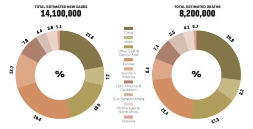
Figure 1 which indicates total number of cancer cases and deaths globally in 2012.
From a personal standpoint, cancer has touched the lives of many of our loved ones.
Tony’s Story: My Nana was diagnosed with renal cancer when she was in her sixties. Through treatment and healthy living, she lived to be 87 years old. Her oldest daughter, my Tita Gilda, was diagnosed with renal cancer two years ago and underwent an operation that removed the malignant mass from her kidney. A malignant tumor is cancerous because abnormal cells divide uncontrollably and can invade nearby tissues. After a successful surgery, she is now in remission, which means she has recovered temporarily.
Tommy’s Story: My great grandmother, Mammoo, was diagnosed with lung cancer in her seventies, and did not want cancer treatment. Unfortunately, she died at 84 years old from lung cancer, and thus greatly affected those around her, including myself.

 (Left) Tommy with his great-grandmother, Mammoo. (Right) Tony with his Nana, Lulu.
From these stories, we only understood cancer at a superficial and personal level. Thus, we decided to take Advanced Biology this year in order to answer the abundant amount of questions we had regarding the human body. Over the past weeks, we have tackled DNA structure and replication, the cell cycle, mitosis and cytokinesis, and the control of the cell cycle. We were amazed to find out how such processes are connected, regulated, and are integral to the development of cancer.
Cancer is the uncontrolled proliferation of cells in the body. Since cancer arises naturally from one’s own body, there is some sort of transition from a normal process to an aberrant process. To best understand this transition, it is important to understand how cells are regulated in a normal and healthy organism in contrast to what occurs when cancer arises.
The body is composed of specialized tissues. These tissues are made up of cells, the smallest unit of life. The collection of cells plays an important role in the body’s systems. For instance, cells are responsible for converting sugar to useable energy, and for receiving signals from our outside world that stimulate our sense of vision and olfaction. You may wonder, how does each cell know its role? Each cell has its own blueprint. This instructor’s manual is known as deoxyribonucleic acid, or DNA (Figure 2). DNA is double helix structure made up molecules called nucleotides. To create the double helix structure, these nucleotides must pair. The nucleotide base called adenine pairs with thymine, and guanine pairs with cytosine. The sequence of the nucleotides ultimately determines the information available for building an organism. Each person, aside from identical twins, has a unique DNA code. It is this code that tells the body’s cells what to do through the production of proteins.

Figure 2 depicts DNA structure and the 4 nitrogenous bases.
There are trillions of cells in the human body. Each type of cell has different life spans, just like different organisms. The body needs to replenish its cell populations. The cell cycle describes the series of events that occur when a cell is replicating. The four main phases are: Gap 1, Synthesis, Gap 2, and Mitosis (Figure 3).

Figure 3 depicts the four phases of the cell cycle along with their checkpoints.
In Gap 1, the cell grows and performs its assigned function. For example, a skin cell grows and is in charge of holding in important nutrients and fluids. Before the cell proceeds to the next phase, it must pass the Gap 1 checkpoint. At this checkpoint, the cell is examined to see if it is large enough to divide and the DNA is examined to see if it remained intact.
After the cell clears the Gap 1 checkpoint it enters the Synthesis phase of the cell cycle. To produce two similar daughter cells, the complete DNA instructions in the cell must be duplicated through DNA replication. Again, the cell must pass the synthesis checkpoint before advancing to Gap 2. If there is an error in DNA replication, a tumor suppressor gene halts the cell cycle, activates DNA repair genes, and fixes the error. Like in Gap 1, if the damage is too excessive, the cell will be programmed to undergo apoptosis, or cell suicide.
Once DNA is replicated and passes the Synthesis checkpoint, it advances to Gap 2. During Gap 2, the cell continues to grow and produce more proteins that are necessary for cell division. As in Gap 1, the cell must pass the Gap 2 checkpoint. Tumor suppressor genes again check for sufficient growth as well as errors in the DNA. If excessive damage is found, apoptosis will occur.

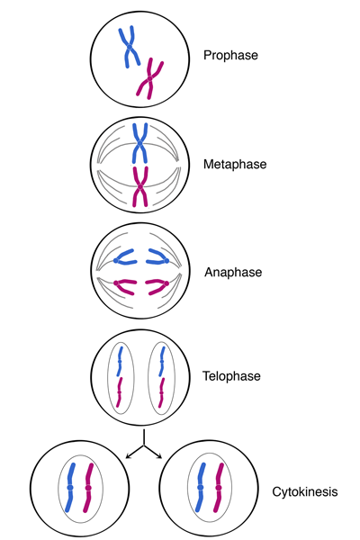
Figure 4 displays the processes of mitosis and cytokinesis. The cell begins to create two nuclei, and cytokinesis results in two daughter cells.
The cell is now is able to proceed to the final phases of the cycle: mitosis and cytokinesis. Mitosis is the division of the nucleus and consists of 5 phases: prophase, prometaphase, metaphase, anaphase, and telophase (Figure 4). These phases systematically divide the copied DNA into two equal sets. Prior to anaphase, the cell reaches the Mitosis checkpoint, which ensures that the cell is dividing properly. If so, the cell continues to divide. When there is an error in cell division, an inhibitor protein prevents the cell from advancing to anaphase. In the process of Cytokinesis, the cytoplasm of the cell divides, resulting in two new cells. This entire process is integral to life. When mitosis and cytokinesis do not occur properly, cell division cannot occur or cells are produced that will not function properly.
Checkpoint Regulators:
The proteins described at each checkpoint are called “checkpoint proteins” or “cell cycle regulators.” These proteins regulate the progression from one phase of the cell cycle to the next. The progression through these checkpoints is a process, which should occur without errors. When errors do occur, the consequences can be catastrophic, and may result in cancer.
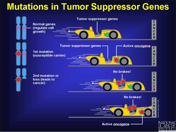
There are two classes of regulators: proto-oncogenes and tumor suppressor genes. Proto-oncogenes are positive regulators of the cell cycle; tumor suppressor genes are negative regulators. One way to think about these regulators is as the accelerator and brake of a car. Similar to the way a car functions through the combination of an accelerator and brake, the growth of the cell is controlled through the proto-oncogenes which accelerate growth, and the tumor suppressor genes that slow cell growth. For each gene there are two copies: one from the mom and one from the dad. In order for a car to function properly, the accelerator and brake must also be functioning properly. When mutations occur in these genes, the cell is unable to function properly. A mutated proto-oncogene results in an oncogene. When activated, oncogenes are like an accelerator that is pushed and stuck to the floor; the cell begins to grow uncontrollably. Oncogenes are a dominant mutation, and a single copy of an oncogene is sufficient for expression of the mutated protein. This means that a single oncogene has the ability to cause an alteration in the growth of a cell. If a car had two accelerators, and one of them is pushed to the floor but the other isn’t, the car would still speed up. Although oncogenes alter the growth of a cell, a single oncogene does not have the ability to cause cancer. The rate of cell division will increase, and future mutations may be more likely, but the brakes of the cell are still functioning and will work to slow division. However, if a tumor suppressor gene becomes mutated, these genes are unable to stall the cell cycle when there is an error. Unlike oncogenes, one single mutated tumor suppressor gene is unable to alter the expression of the growth trait, and is a recessive gene. If a car had two brakes, and one of the breaks was defective, the car could still stop due to the other brake. When there is a combination of mutated proto-oncogenes and tumor suppressor genes in the same cell, uncontrollable cell division occurs, resulting in a higher probability for cancer to arise (Figure 5). For example, if the DNA is damaged, a tumor suppressor gene called p53 determines whether the cell will commit apoptosis. Additionally, p53 is a transcription factor which transcribes, or converts, DNA into RNA. p53 enters the nucleus and transcribes p21 to stall the cell cycle. When P21 is present, it stalls the cell cycle until the DNA is repaired. P21 is able to do this because it is an allosteric inhibitor (Figure 4). P21 binds to the inhibition site of Cyclin Dependent Kinase. An allosteric inhibitor is a regulator molecule, which binds to an enzyme (CDK). Then, it causes a conformational change, which alters the active site of the enzyme. Thus, the molecule cannot to bind to the active site of the enzyme. In this case, cyclin is not able to bind to the enzyme, Cyclin Dependent Kinase (CDK). Without the binding between Cyclin and CDK, the cell cycle stops, and the only way for the process to continue is based upon the concentration levels of cyclin.

Figure 5 displays the way an allosteric inhibitor works.

Figure 6 depicts the mutations in proto-oncogenes and tumor suppressor genes and how they affect cancer.
How does cancer arise?
When the genetic “blueprint” of DNA is changed or mutated, cancer may arise. Carcinogens are substances to which humans are exposed that can cause cells to divide at a faster rate, which then can alter DNA, leading to cancer. Lifestyle factors like using tobacco, naturally occurring exposures such as ultraviolet light, medical treatments such as radiation, workplace exposures, and pollution are examples of carcinogens that have the ability to mutate genes. Mutations can be inherited from parents as well. A mutation can also be caused through a mis-copying during mitosis when the nitrogenous base pairs match up incorrectly (Figures 7 and 8).

Figure 7 displays DNA of a normal cell with base pairs matching up correctly.

Figure 8 depicts this DNA as mutated and one of the base pairs does not match up.
Mutations and p53:
Unregulated cell division causes the mutations to spread. When there is a defective tumor suppressor gene, especially p53, a cell with damaged DNA may proceed through the stages of the cycle, ultimately dividing. The daughter cells will inherit more mutations due to the unrepaired DNA of the mother cell through the division of cells. Cells with defective p53 tend to accumulate mutations. The function of p53 is to detect mutations and make the cell fix the mutations, or cause the cell to undergo apoptosis. As a result, proto-oncogenes can become oncogenes or deactivate other tumor suppressor genes within the cells, thus passing on damaged DNA (Figure 9).

Figure 9 displays a cell accumulating mutations and growing uncontrollably.

Figure 10 displays the process of how a mutated p53 affects the regulation of the cell cycle.
Dividing Cells:
As cells continue to divide, there is a higher chance that some of the cells may have mutated further. Some cells will contain multiple mutations and duplicate at a faster rate. As more mutations appear through the increase in cell division, the growth of a tumor, known as an abnormal amount of cells, will increase. This tumor is currently contained within the tissue. This type of tumor is a benign tumor, and is not yet dangerous.
Following more uncontrolled division, mutated cells are able to invade the membrane of the tissue. Angiogenesis is the formation of new blood vessels that supply the cancer cells with oxygen and nutrients. This supply allows cells to divide more uncontrollably because they are not as dependent on the normally functioning systems. Angiogenesis results in a rapid increase in the size of the tumor, as the number of cancer cells approach a billion. Cancer cells have the ability to metastasize, or migrate to other parts of the body. Cancer cells enter the blood vessels, which form due to angiogenesis, and travel to other parts of the body via the bloodstream (Figure 10). To metastasize at a new location, a tumor cell needs to exit the vessel system and invade new tissue. The cell must attach itself to a vessel’s wall, and work its way through the vessel and enter the tissue. This tumor then becomes malignant, as it is dividing rapidly and able to invade other tissue, unlike a benign tumor, in which the tumor remains in the same tissue. Therefore, cancer mutations are deadly because they eventually can spread to the entire body.
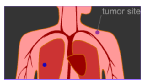
Figure 11 depicts the cancerous cells traveling through the bloodstream to other parts of the body.
A cause worth helping:
Cancer, a collection of abnormal cells, does not discriminate. It affects many populations around the world. After many years of research, scientists and doctors have still not been able to find a complete cure for this disease, and it is unclear if there ever will be one. However, there have been major cancer breakthroughs, which give us hope that this unyielding disease will soon be annihilated. As students, we are responsible for comprehending new concepts and educating others. During this project, we delved into the basic parts of cancer and hope to present this information to others on a diverse set of platforms. Additionally, we discovered how vital it is to recognize the human side of science, which we were able to do by connecting multiple concepts we learned in class to the hardships our families have faced due to cancer. With social media today, we are able to reach millions of people from across the globe and possibly spark an individual to join us in the fight against cancer.
 Tony Banson is V Form day student from Hopkinton, MA. Tommy MacNeil is a V Form boarding student from Lancaster, MA.
References
Cancer and the cell cycle [Image]. (2015). Retrieved from
Cancer Facts & Figures 2017. (2017). Retrieved May 9, 2017, from American Cancer Society
Cancer is a leading cause of death worldwide [Image]. (2012). Retrieved from
http://canceratlas.cancer.org/the-burden/Chris. (2016, January 27).
Allosteric inhibition [Image]. Retrieved from
DNA Structure [Image]. (n.d.). Retrieved from http://visual.ly/dna-structure.
The Eukaryotic Cell Cycle and Cancer. (2014, November 21). Retrieved May 9, 2017, from
Biointeractive website: http://www.hhmi.org/biointeractive/eukaryotic-cell-cycle-and-cancer
Interphase. (n.d.). Retrieved May 9, 2017, from Education Seattlepi website:
http://education.seattlepi.com/happens-sphase-interphase-5508.html
Lodish H, Berk A, Zipursky SL, et al. Molecular Cell Biology. 4th edition. New York: W. H.
Freeman; 2000. Section 24.5, Mutations Affecting Genome Stability. Available from: https://www.ncbi.nlm.nih.gov/books/NBK21551/
Metastasis [Image]. (2001, February). Retrieved from
http://www.pbs.org/wgbh/nova/cancer/grow_nf11.html
Mitosis [Image]. (n.d.). Retrieved from
http://cyberbridge.mcb.harvard.edu/images/mitosis5_1.png
Oncogenes and Cancer [Image]. (2013, January 11). Retrieved from
https://oncogenesandcancer.files.wordpress.com/2013/11/cancer53.jpg
Statistics for Different Kinds of Cancer. (2016, August 16). Retrieved May 9, 2017, from Centers
for Disease Control and Prevention website: https://www.cdc.gov/cancer/dcpc/data/types.htm


