
Home » Posts tagged 'STEM Fellowship'
Tag Archives: STEM Fellowship
Inverted Airfoils’ Abilities to Prevent Wind Storm Roof Destruction
By Yolanda Zhou, VI Form
Inverted Airfoils’ Abilities to Prevent Wind Storm Roof Destruction
Student-Submitted Note: This paper investigates a new way to reduce aerodynamic lift created around houses’ roofs in extreme windstorms. Three test models were created in computer modeling software OnShape and simulated in CFD using OpenFOAM. This project was inspired by the concept of “downforce” used to stabilize Formula One race cars. This paper was written for submission to the High School Science Symposium (HiSci) in May 2023 as part of the STEM Fellowship.
Abstract
As severe windstorms increase in frequency and intensity, more residential houses are predicted to be vulnerable to structural damage by severe windstorms towards the end of the 21st century. Wind around the roof exerts a negative pressure that lifts the roof up and threatens the structural integrity of houses. In this paper, a novel method of mitigating lift using inverted airfoils was explored through CFD simulations. The performance of the model was assessed through three criteria: net lift coefficient, effectiveness under different wind directions, and manufacturability. Visual representations of pressure and velocity distribution of the airflow over the model were analyzed to validate simulation data. Single inverted airfoils and round airfoil-shaped roofs were tested for the first prototype. A further iteration of the prototypes improved the model performance. All roof configurations were able to reduce the overall lift of the system when compared to the control group. Single airfoil roof setup was most effective at reducing net lift, while round roof setup was effective in a wider range of wind direction conditions. Rectangular Roof setup combines the advantages of both setups and reduces more net lift than the Revolved Airfoil setup.
Introduction
As global climate change accelerates, the intensity and frequency of wind disasters, such as tropical cyclones and tornadoes, are predicted to increase significantly by the end of 21st century [8]. The increase in the intensity of tropical cyclones is found to be correlated to the rise of Sea Surface Temperatures [8]. Furthermore, Category 4 and 5 tropical cyclone activities, which have wind speeds that exceed 150 miles per hour, are likely to increase in the late 21st century [10]. Such tropical cyclones, although very rare, are catastrophic and account for around half of the economic damage done by all tropical cyclones in the US [6]. In 2013, an EF-5 category tornado hit Moore, Oklahoma. Reaching a speed of over 200 miles per hour, type EF-5 tornadoes are the rarest and most destructive type of tornado on Earth. The tornado caused 24 deaths, more than 200 injuries, and billions of dollars for repairing the destroyed houses and facilities [3]. Overall, tropical cyclone frequencies, intensities, and damages are projected to increase as the global climate continues to get warmer [10]. High School Science Symposium 2022
(more…)
The Impact of Black Sesame Pigment on Drosophila Melanogaster with Alzheimer’s Disease
By Ally Bauer, VI Form
The Impact of Black Sesame Pigment on Drosophila Melanogaster with Alzheimer’s Disease
My project for the STEM Fellowship was studying the impact of black sesame pigment on drosophila melanogaster with Alzheimer’s disease. Alzheimer’s Disease is a neurodegenerative disease that affects over 44 million people worldwide. One of the hallmarks of Alzheimer’s Disease are amyloid-beta plaques that form in between the neurons and disrupt cell communication and function. Black sesame pigment, derived from black sesame seeds, has been proven to reduce the aggregation of these plaques in vitro studies. Drosophila melanogaster, or fruit flies, are wonderful model organisms that are utilized for their quick reproduction rate, easily manipulated genome, and the relation its genome has with humans. For my experiment, I was able to track the progression of memory loss in flies with Alzheimer’s disease. I had multiple groups to show me if greater concentrations of black sesame pigment would slow the progression of memory loss in the flies. Although the results of the experiment proved my null hypothesis to be correct, I now have a greater understanding of Alzheimer’s Disease, the scientific method, and having control over what I learn and how I learn it. I am incredibly grateful for Ms. Lohwater, Mr. Loomer, Mr. Valitutto, and the five other STEM Fellows who have mentored me, shaped my project, and problem solve throughout the year.

Bio-ink: Evaluation of Protein as Biomaterials for 3D Bioprinting
By Jiwon Choi, VI Form
Editor’s Note: This STEM Fellowship project by Jiwon won the Worcester Regional Science and Engineering Fair (1st out of 130+ students). She placed third out of all 200+ projects at the Massachusetts Science and Engineering Fair allowing her to compete at 2019 ISEF in Phoenix.
Three-dimensional (3D) bioprinting is one of the most promising methods of tissue engineering as it provides unprecedented versatility and precision in delivering cells and biomaterials to desirable places. However, limitations still exist in the availability of bioinks with natural bio-macromolecular components. In this research, chicken albumin is evaluated as a potential bioink for direct extrusion bioprinting of hollow constructs through alginate-templated crosslinking. Channel diameter, wall thickness, and bioink feed rates are calculated to assess the printing performance of the alginate-based bioink. It is shown that an albumin-based bioink with as low as 1.33% of total alginate concentration can be employed to successfully print microfibrous hollow constructs with a uniform diameter.
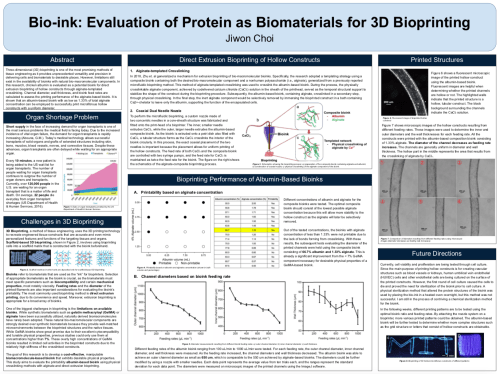
Click on Image to View PDF of Jiwon’s Poster

The Effects of the Ketogenic Diet’s Effect in a Drosophila Melanogaster Model of Parkinson’s Disease
By Grant Gattuso, VI Form
The Effects of the Ketogenic Diet’s Effect in a Drosophila Melanogaster Model of Parkinson’s Disease
Abstract
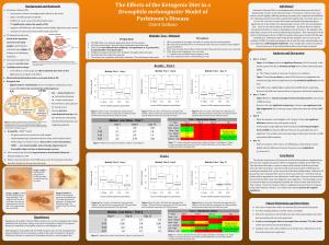
Click on Image to View PDF of Poster
Parkinson’s Disease (PD) is a neurodegenerative disease that causes the loss of dopaminergic neurons in the substantia nigra. This disease is chronic and causes tremors, muscle rigidity, difficulty speaking, and many other symptoms that debilitate the individual and deteriorate their quality of life significantly. Currently, there is no cure for PD. Previous research shows that mitochondrial dysfunction plays a significant role in the death of the dopaminergic neurons in PD. Since the ketogenic diet – a four to one ratio of lipids to carbohydrates – has been shown to improve mitochondrial function in diseases like Epilepsy and Alzheimer’s, the ketogenic diet could delay or improve the onset of Parkinsonian symptoms. This study measured the effects of the ketogenic diet in a PINK1Drosophila melanogaster model of PD through a mobility test. Preliminary data found that the ketogenic diet can increase the mobility of PINK1 Drosophila melanogaster for at least four days and potentially even up to eight days, confirming the hypothesis. Four to eight days could correspond to many human years if the same beneficial effects were found in humans.
To read Grant’s full STEM Fellowship paper, click here. (more…)

Unique Advances in Transplant Research with Hydractinia
By Haley Dion, VI Form
Unique Advances in Transplant Research with Hydractinia
Transplantation is the future of medicine. It is an ever-evolving field of research. For three weeks this summer, I was given the opportunity to take part in the research by interning at the Thomas E. Starzl Transplant Institute. At the institute, I worked in the Nicotra Lab under the mentorship of Dr. Matthew Nicotra. The Nicotra Lab is one of the Stuart K. Patrick Research Laboratories at the Institute named after St. Mark’s alumnus, Stuart K. Patrick ’57. The lab I worked in is unique because it works with an organism that is very rarely used in research: Hydractinia

Hydractinia are invertebrates that live on hermit crab shells. These organisms are part of the cnidarian species, and they grow as colonies. Hydractinia grow mat tissue, which is the base of their colony. Within the mat, there are gastrovascular canals that allow cells to flow throughout the colony. Some Hydractinia have stolons, branched stem-like structures, that extend from their mat. Hydractinia also have polyps that protrude from the top of their mat. These polyps are tubes surrounded by tentacles that are used to consume food. In addition to the polyps that help the Hydractinia eat, there are reproductive polyps that can be used to tell whether the colony is male or female. This image illustrates the development of a Hydractinia embryo to a colony. The image shows what an adult polyp looks like, in addition to both the male and female sexual polyps. (more…)

Software Pipeline Connecting Close-Range Photogrammetry and 3D Printing
By Gillian Yue, VI Form
Software Pipeline Connecting Close-Range Photogrammetry and 3D Printing
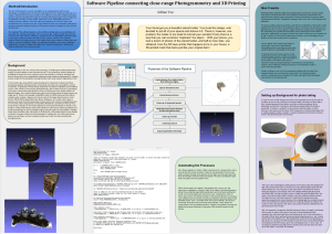
Click on Image to View Full Poster
Abstract/Introduction
The aim of this project is to make it possible for an average person with no prior knowledge in photogrammetry to 3D-print small objects found in daily lives. My work is to create a software that serves as a pipeline; the software connects the multiple processes that are required to transform the input of of photos of the target object into an output of a 3D printable model file. In other words, what used to be a complicated process of switching between different tools and manually processing the model to make it 3D printable becomes a simple one-click routine where the user can provide the initial group of photos, and then simply sit next to the 3D printer to wait for the object to come out half an hour later. (more…)